

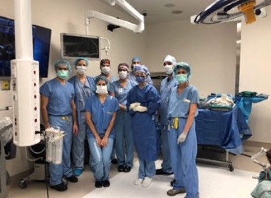
By: Salvatore Di Maio, MD CM, FRCSC (neurosurgeon); Tamara Mijovic MD, CM, FRCSC (otolaryngologist); Diane Bouchard (medical electrophysiology technician); Paul Wieczorek MD, (anesthesiologist), Jewish General Hospital, Montreal, Quebec
The Acoustic Neuroma Association of Canada has been a great resource tool for our patients, for the support found from connecting online and in-person with other patients with the same issues, and also for understanding the various considerations in making medical decisions, and what different treatments actually entail from a patient perspective. One topic of interest which recently came upon one such group is how hearing and facial motor function are monitored during surgery. This is referring to Intra-operative monitoring (IOM), i.e., the various techniques used to observe and monitor neurological functions during surgery. The purpose of this article is to understand how the surgical team monitors neurological functions using IOM while patients are undergoing acoustic neuroma surgery. It’s a great topic from our perspective too because it really highlights the fact that not just IOM but the whole acoustic neuroma operation is really an orchestrated feat made possible through teamwork. This is why the article is the shared perspective and contribution of the members of the surgical team, including the neurosurgeon, otolaryngologist, anesthesiologist, and medical electrophysiology technician.
There is no doubt that surgery is an important treatment option for patients with acoustic neuromas, but it is also invasive and invariably there are risks involved. While a lot of the success of surgery ultimately has to do with the capacities and experience of individual surgeons, certain techniques such as IOM help make surgery safer and more successful. The use and technique of intra-operative monitoring are not unique to one centre for the most part, and we would imagine that most surgeons that operate routinely on patients with acoustic neuromas would implement some version of the techniques grossly described here. Indeed, most otolaryngologists would consider this the standard of care.
For IOM, a technologist or neurophysiologist uses electrodes (tiny needles) and computers to continually assess the function of the nervous system throughout the whole surgery. For acoustic neuroma surgery, the main functions we are interested in is 1) facial nerve/motor function and 2) hearing, which are further described below, although there are several other neurological functions we can monitor (including during acoustic neuroma surgery) but that won’t be touched on here. We are getting real-time feedback about the state of facial and auditory nerve function, and with stimulating instruments we can use IOM to help locate certain structures (e.g., the facial nerve) which can sometimes otherwise be hard to locate visually when compressed by the tumour. When either irritation of a nerve or a decrease in its function is detected, it can provide the surgeon with early warning and ideally enough time to respond and act accordingly before permanent injury occurs.
Facial Nerve Monitoring
Practically speaking, facial nerve monitoring involves the placement of an electrode inside a muscle of the face that is innervated by the facial nerve and records the electrical activity generated by the stimulation of that muscle. Most frequently, we place at least two electrodes in the facial muscles on the same side of the tumour: one at the level of the eye (orbicularis oculi muscle; the muscle that closes the eyelid) and one at the level of the lip (orbicularis oris muscle, which animates the mouth) as these are branches far away one from the other that happen to both provide vital facial nerve functions.
When the facial nerve is stretched by dissection, heated by the cautery or stimulated by a stimulating instrument, it creates activity that is transmitted to the muscles that it innervates and the muscle contracts. The contraction is picked up by the electrode that is connected to a monitor. Most monitors assign a sound to the contraction and a noise is generated by the machine that signals to the surgeon in real-time that the facial nerve is being stimulated. We use this information together with anatomical knowledge, clinical judgement, and technical skills to remove all of the tumour safely or to leave a small remnant on the facial nerve in some cases to preserve its function.
Besides helping surgeons locate the nerve and avoid trauma, monitoring the nerve function provides prognosis on outcome after surgery. It can inform us about the integrity of the nerve and the likelihood of function recovery. This means that so long as certain thresholds in IOM are maintained, a patient who wakes up after surgery with facial weakness will nevertheless go on to recover function over time. The data basically helps us understand if the nerve is temporarily stunned by all the surgical work but otherwise intact, versus irreversibly damaged. This is why the rate of initial facial nerve weakness after surgery is around 10-15% but permanent facial nerve paralysis is a lot rarer.
Auditory Function Monitoring
Many patients with acoustic neuromas will already have experienced hearing loss by the time the tumour is even diagnosed, and currently it is generally not possible to restore hearing function even if the tumour is removed. There are, however, a proportion of patients who still have functional hearing going into surgery, and in those patients, hearing preservation is an important goal.
In general, monitoring of cochlear nerve function is more challenging than facial nerve monitoring. One reason is that hearing is technically a lot harder to assess in a patient that is asleep. Another reason is that hearing not only depends on the integrity of the cochlea, the cochlear nerves and the brain pathways that analyze sound, but is also very sensitive to the fluctuations in blood supply to these structures which can occur during the surgical procedure.
The most common method of intraoperative auditory monitoring is through Auditory Brainstem Responses (ABRs). For this monitoring to work, the patient needs to have good hearing before surgery and a good ABR at baseline. Most often, the tumour has already stretched the cochlear nerve enough that we are unable to get an ABR and the monitoring is not possible. The cochlear nerve is very thin and splayed out at its entrance into the cochlea which makes it more vulnerable. This also at least partially explains why a lot of patients with acoustic neuromas present initially with a complaint of not hearing as well from one ear in the first place. The other challenge is that auditory monitoring does not provide instantaneous real-time and direct information the way facial nerve monitoring does. In auditory monitoring, there is always a lag, meaning that what is seen on the monitor is the impact from the surgical move that was done several seconds before. The window to identify and then react to problems in auditory monitoring is smaller, and so from a surgical standpoint hearing preservation rates are generally much lower than with facial nerve preservation, particularly for larger tumours.
The technique of ABR involves placing an occlusive earphone in the ear canal at the start of surgery. This earphone emits sounds and clicks that are transmitted through the ear canal to the eardrum, through the ossicles and finally to the cochlea. The cochlear nerve is stimulated by the noise and the resultant electrical activity is sent through the nerve to the brainstem and the cerebral cortex. Electrodes in the scalp collect these signals and the monitor will decipher a signature tracing (i.e., the ABR) by averaging out any other electrical noise through repetition and averaging over time. The amplitude (size) and overall timing of the ABR can tell us about the health of the cochlear nerve as well as the brainstem and brain pathways they relay to. Because the monitor depends on the average of hundreds of ABRs (albeit over very short periods of time), when changes are noted, some permanent cochlear nerve damage may already have occurred.
Anesthesia
The anesthesiologist and anesthesia team will ensure that patients are asleep and comfortable during the entire operation. Some of the standard medications that are used for anesthesia can, however, interfere with the ability of the electrophysiology technician and surgeon to appropriately monitor the nerves as described above. The anesthesiologist caring for these patients must therefore select alternative medications that will not only ensure the patient is unconscious, but minimizes interference with the intra-operative monitoring. For instance, in order for the surgical team to monitor the function of the facial nerve, the anesthesiologist must ensure that the patient’s muscles are not weak and not paralyzed. Similarly, the electrical signals in the nerves being monitored can be diminished with certain anesthetic medications, and alternatives must be selected (often in the form of intravenous anesthesia). The anesthesiologist, electrophysiology technician, and surgeon are in constant communication during the surgery to ensure good intra-operative monitoring signals are obtained, and the anesthesiologist can adjust the anesthetic regimen to help obtain good electrical signals, all while ensuring the patient is safe and asleep.
Conclusion
As a team, we are always looking for ways to make treatment for acoustic neuromas safer. Intra-operative monitoring is one technique we use in surgery to improve outcomes for facial and hearing nerve function. For anyone who wants to read more about this, a simple Google search for “intra-operative monitoring” and “patient information” yields plenty of well-written descriptions.